Neuroelectric and Hemodynamic Correlates of Śūnyatā Meditation – Combined fMRI-EEG Study
RANGANATHA SITARAM 1, MICHAEL ERB 2, ADRIAN FURDEA 1,
QUANG CHIEU NGUYEN VAN HUNG 3, MASTER THICH THONG TRIET 4
1 Institute of Medical Psychology and Behavioral Neurobiology,
University of Tübingen, Germany
2 Section Experimental MR of the CNS, Department of Neuroradiology,
University Hospital, Tuebingen, Germany
3 Śūnyatā Meditation Stuttgart e.V., Germany
4 Śūnyatā Meditation Center, Perris, CA, USA
Corresponding author: sitaram.ranganatha@uni-tuebingen.de
Note: this article was displayed at the Annual Meeting of the Organization for Human Brain Mapping (OHBM) in 2010 in Barcelona, Spain
INTRODUCTION
The word meditation describes practices that self-regulate the body and mind. Śūnyatā (Sanskrit – emptiness) meditation stems from the Buddhist philosophy that signifies the impermanent nature of form; meaning that objects in the world do not possess essential or enduring properties. In Buddhist spiritual teaching, cultivating insight into emptiness leads to wisdom and inner peace. Śūnyatā meditation practice is aimed to develop an ability to avoid discursive (wandering, long-winded) thought, and instead acquire insight into the nature of reality through direct perception of the internal (bodily) and external (sensory) states.
AIM and HYPOTHESES
The aim of the present study was to investigate state changes in the brain and physiology during Śūnyatā meditation when participants are confronted with a variety of external stimuli: visual, auditory and tactile. Based on the rationale behind the Śūnyatā practice, we hypothesized that the following state changes occur during meditation in comparison to normal day-to-day thinking:
- Memory retrieval, planning and executive control areas of the brain will be deactivated;
- Brain areas related to interoception and sensory perception will be activated.
MATERIALS and METHODS
Participants: Experienced Meditators
Participants included the Śūnyatā meditation master Thích Thông Triệt (age: 80 years, years of meditation experience > 30, meditates 7 hours per day), 1 other expert monk and 2 expert nuns (mean age: 70 years, years of meditation experience > 15, meditate 4 hours per day), and 2 more intermediate practitioners (mean age: 50 years, years of meditation experience > 5, meditate 1 hours per day).
Experimental Protocol
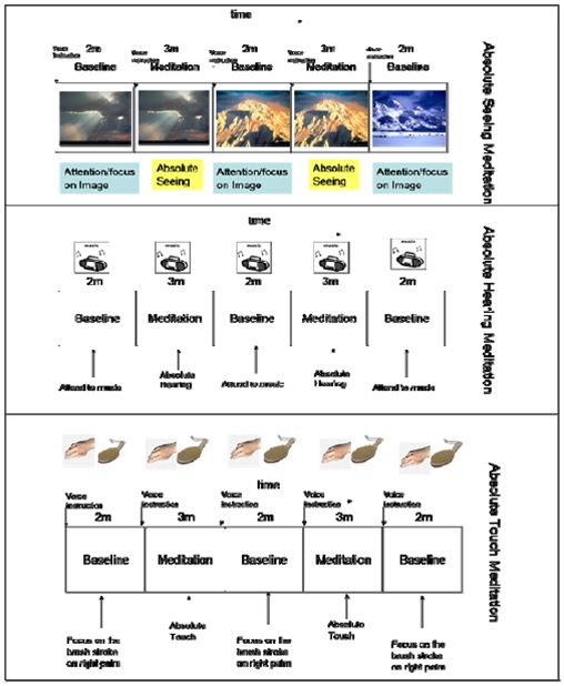
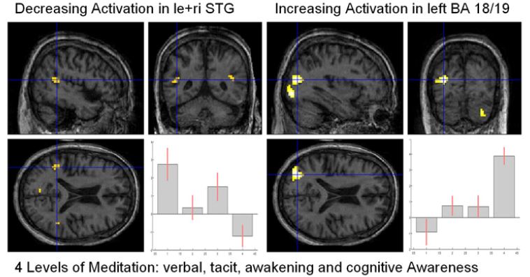
The experimental protocol comprised of 3 blocks of baseline, normal day-to-day discursive thinking (duration=2min) and 2 blocks of meditation (duration=3min) alternating with one another. Four methods of meditation, namely, absolute seeing, absolute hearing, absolute touch and absolute cognition were measured in separate sessions, with the above protocol, in accompaniment with visual, auditory, touch stimuli and no stimuli, respectively. Identical stimuli were presented in the baseline and meditation blocks to tease-out the influence of meditation practice on brain activations. In addition, four different levels of meditation depths as described in Buddhism were investigated, namely:
- Verbal awareness
- Tacit awareness,
- Awakening awareness
- Cognitive awareness.
These measurements were performed in the same block design as above.
fMRI, EEG and Physiological Measurements(electroencephalogram)
RESULTS
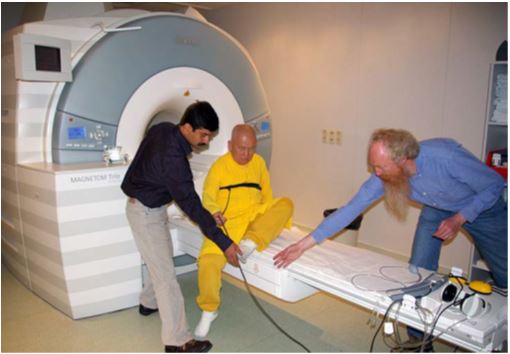
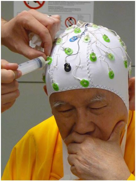
A standard echo-planar imaging (EPI) sequence on a 3T whole body scanner (Siemens, Erlangen, Germany) with the following parameters was used: TR=3000ms, TE=40ms, number of slices=36. For superposition of functional maps upon brain anatomy a high-resolution T1-weighted structural scan of the whole brain was collected from each subject. The above protocols were repeated on different days (5) in a series of measurements spanning 3 years, with the last session utilized for simultaneous measurement of EEG (with BrainAmps MR compatible amplifier and EEG cap) and fMRI. In addition, pulse and respiration were measured and at the end of each run, meditators were requested to make subjective ratings of depth of meditation they achieved in each block of meditation.
fMRI Results
Group Independent Component Analysis Results
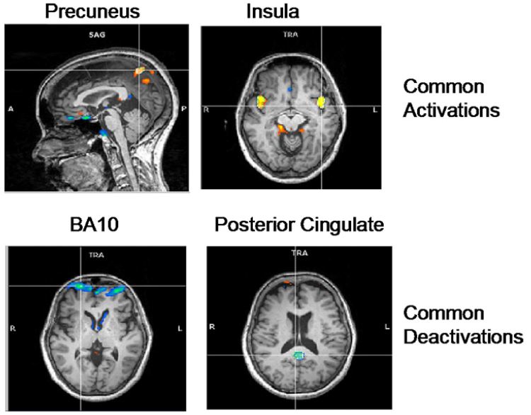
Common activations and deactivations across meditation types:
- Activation in bilateral Precuneus, implicated in self-processing and consciousness (Cavanna, 2007).
- Activation in the bilateral insula, implicated in interoception (Craig, 2008).
- Deactivation in frontopolar region of the brain, namely, BA-10, involved in strategic processes including memory retrieval and executive function.
- Deactivation in the Posterior Cingulate, implicated consistently in the default network of brain function (Raichle, 2000).
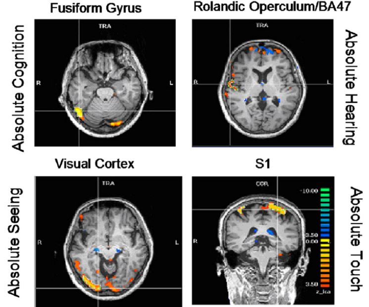
Meditation type specific activations:
- Enhanced activation of the fusiform gyrus (FFG) during absolute cognition meditation,
- Enhanced activation of the right rolandic operculum and inferior frontal gyrus (BA 47) during the absolute hearing meditation,
- Enhanced activation of the visual cortex during the absolute seeing meditation, and
- Enhanced somato sensation during the absolute touch
Analysis of different meditation levels shows a decrease of activations in the left and right superior temporal gyrus (STG) and an increase of activation in the left higher visual areas (BA 18/19), the right inferior frontal gyrus (IFG) and the right insula.
EEG Results
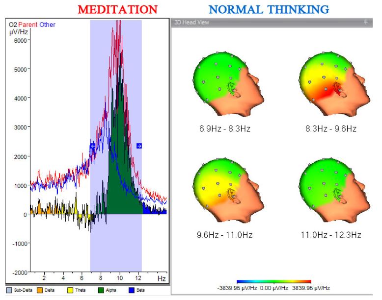
Absolute Seeing Meditation
Meditation of the different senses, namely, absolute seeing, hearing and touch showed increased alpha power in duration meditation compared to normal thinking in the occipital, temporal and sensorimotor regions, respectively, in congruence to the fMRI results.
CONCLUSIONS
Our results show that Śūnyatā meditation enhances perception of external stimuli and interoception (of internal bodily states) as shown by heightened activations in sensory areas and insula when compared to the normal, day-to-day thinking state. This type of meditation reduces discursive thought as shown by a consistent deactivation of the BA-10 involved in memory retrieval, planning and executive function.
References
- Cahn, R., & Polich, J. Meditation States and Traits: EEG, ERP, and Neuroimaging Studies. Psychological
- Austin, J. H. (2006). Zen-Brain Reflections. Reviewing Recent Developments in Meditation and Consciousness. Cambridge, MA: The MIT Press.
![]()