Altered Visual and Auditory Processing in Sunyata Meditation: A combined NIRS and EEG Experiment
1Michael Erb, 2Ann-Christine Ehlis, 2Lena Ernst,
3Van-Hung Nguyen Quang Chieu, 4Master Thich Thong Triet , 2Andreas Fallgatter, 5Ranganatha Sitaram
1Department of Neuroradiology, University Hospital of Tuebingen, Germany,
2Department of Psychiatry and Psychotherapy, University Hospital of Tuebingen, Germany,
3Śūnyatā Meditation Stuttgart, Germany,
4Śūnyatā Meditation Center Perris, Riverside, CA, USA,
5Institute of medical psychology and behavioral neurobiology, University of Tuebingen, Germany,
e-mail: michael.erb@med.uni-tuebingen.de
Note: this article was displayed at the Annual Meeting of the Organization for Human Brain Mapping (OHBM) in 2011 in Québec City, Canada
Introduction
The word meditation describes practices that self-regulate the body and mind. Śūnyatā (emptiness) meditation stems from the Buddhist philosophy that signifies the impermanent nature of form; meaning that objects in the world do not possess essential or enduring properties. Śūnyatā meditation practice is aimed to develop an ability to avoid discursive (wandering, longwinded) thought, and instead acquire insight into the nature of reality through direct perception of the internal (bodily) and external (sensory) states.
The experiment presented here is a continuation of our study described last year (Sitaram et al., 2010) investigating state changes in fMRI and EEG signals during Sunyata meditation.
In the present study, we conducted our experiments with fNIRS as it allows for noise-free measurements and upright, sitting position during meditation.
Questions:
- Is it possible to measure similar effects with fNIRS as with fMRI?
- Is a fNIRS Experiment a more suitabe environment for Meditation than fMRI?
Methods
Subjects:
In the main study 8 participants with different extents of meditation experience (5-30 years) were measured using fMRI and in some sessions with simultaneous fMRI and EEG.
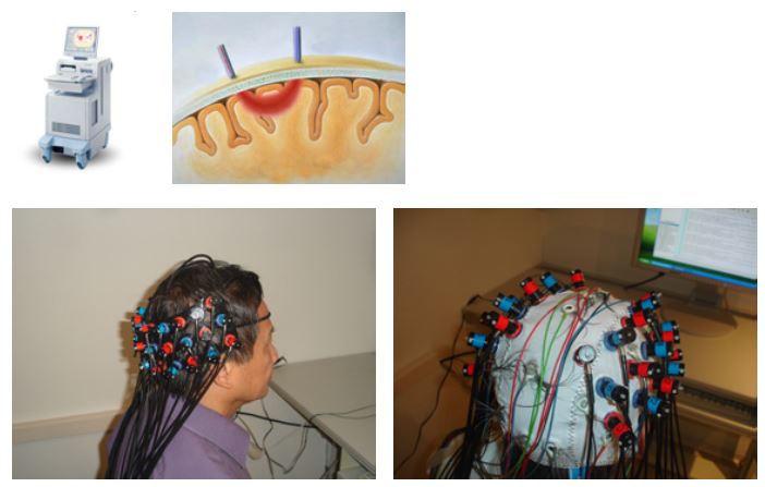
One of the meditators participated in this pilot experiment with combined NIRS (Hitachi ETG-4000, Hitachi Medical Co., Japan) and EEG (24 channel BrainAmp, Brain Products, Germany) measurement (for previous simultaneous NIRS/EEG measurements, cf. Ehlis et al., 2009).
Two configurations of the NIRS optode sets were used: a 3×11 array (52 channels) placed on the occipital pole for visual stimulation on a LED monitor and two 3×5 arrays (44 channels) incorporated in an EEG EASYCAP and covering parieto-frontal parts of the head for auditory stimulation with earphones.
Task design and stimuli:
The experimental protocol comprised 4 blocks of baseline involving normal day-to-day discursive thinking (duration=2min) that were alternated with 3 blocks of meditation (duration=3min).
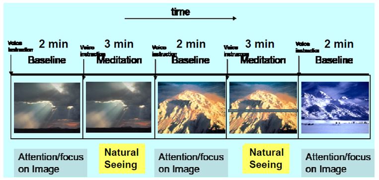
The meditation methods were natural seeing with constant visual stimulation (same pictures for corresponding baseline and meditation blocks) and natural hearing with auditory stimulation (sound of singing bowl every 12s).
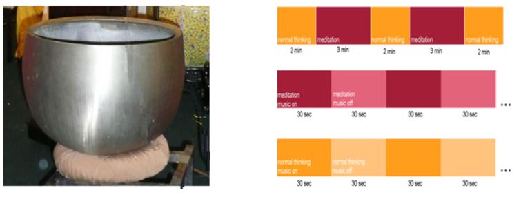
In addition to the tasks with continuous stimulation, 8 alternating shorter on-off blocks of 30s were used, both for continuous normal thinking and continuous meditation.
Data evaluation:
NIRS data were analyzed with SPM5 and the NIRS_SPM toolbox (Ye et al., 2009), the EEG data with SPM8 and the FAST toolbox (Leclercq et al., 2010).
Earlier fMRI & EEG results (OHBM 2010):
Group Independent Component Analysis Results:
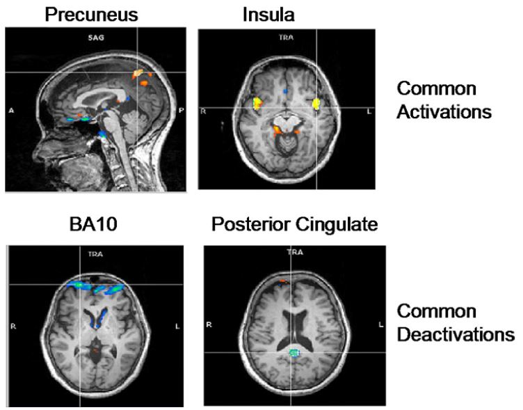
Common activations and deactivations across meditation types:
- Activation in bilateral Precuneus, implicated in self-processing and consciousness
- Activation in the bilateral insula, implicated in interoception
- Deactivation in frontopolar region of the brain, namely, BA-10, involved in strategic processes including memory retrieval and executive function.
- Deactivation in the Posterior Cingulate, implicated consistently in the default network of brain function
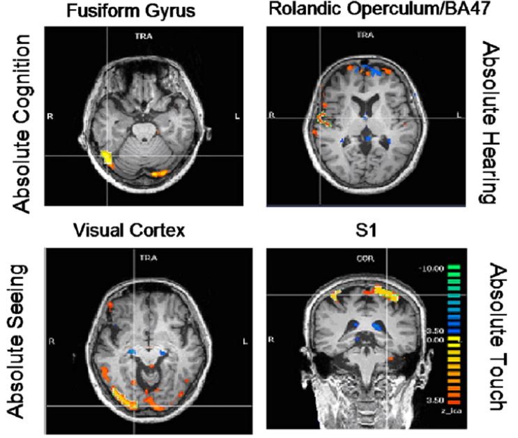
Meditation type specific activations:
- Enhanced activation of the fusiform gyrus (FFG) during absolute cognition meditation,
- Enhanced activation of the right rolandic operculum and inferior frontal gyrus (BA 47) during the absolute hearing meditation,
- Enhanced activation of the visual cortex during the absolute seeing meditation, and
- Enhanced somatosensation during the absolute touch meditation.
EEG Results:
Meditation of the different senses, namely, absolute seeing, hearing and touch showed increased alpha power in duration meditation compared to normal thinking in the occipital, temporal and sensorimotor regions, respectively, in congruence to the fMRI results.
Results
Results of NIRS analysis (see Fig. 1) show enhanced activation in meditation compared to normal thinking with continuous stimulation (1st row) in the primary visual areas (occipital cortex) and parietal regions, with a distinct left-lateralization. Switching stimulation on-off mainly activated the primary visual areas (2nd row, 3rd row) with additional activations in the parieto-lateral regions in the meditation task (3rd row).
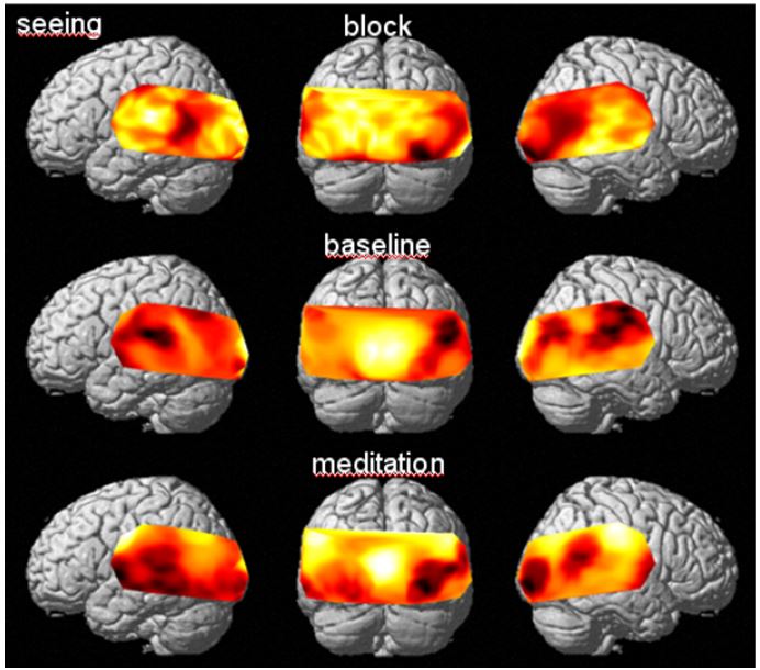
Figure 1: Change of O2Hb concentration with visual task
A similar pattern of activation was found for auditory stimulation (see Fig.2).
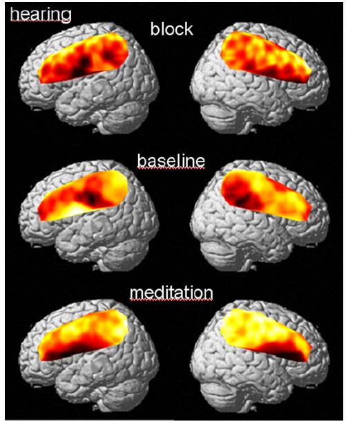
Figure 2: O2Hb Change with auditory task
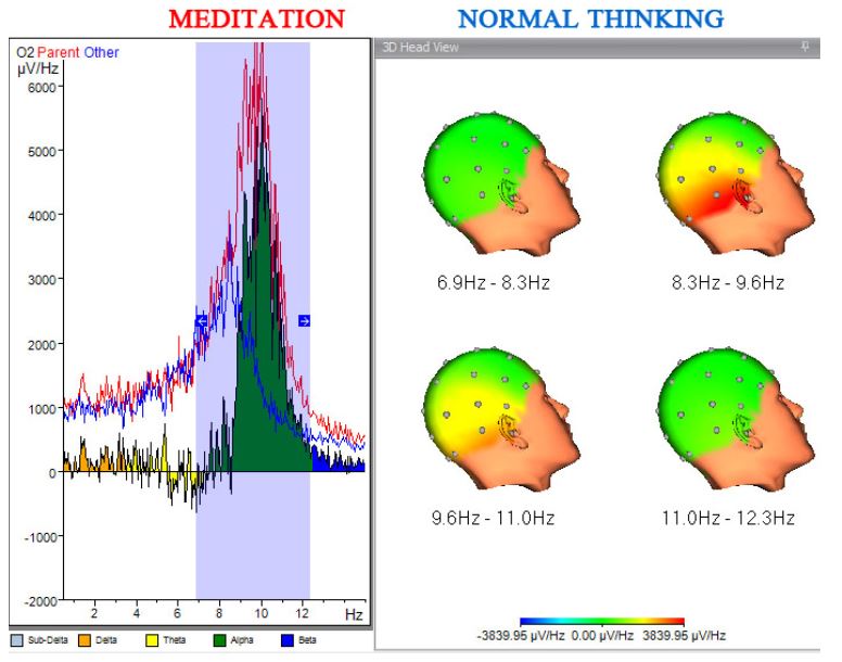
Figure 3 depicts spectra and power in frequency bands from EEG data analysis of the hearing experiments. The block experiment with constant stimulation shows enhanced power in the low frequency bands (delta, theta, alpha) while the participant was in a meditative state (peak at 8.25 Hz: 15 vs. 8.5; peak at 4 Hz: 4 vs. 2).
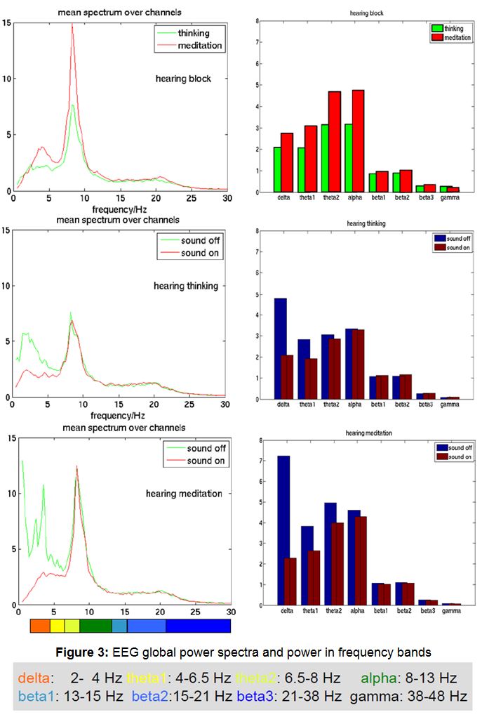
The two runs were auditory stimulation was switched on and off every 30s resulted in an activation peak at 8.25 Hz with a higher value (12) in the meditation condition compared to that of the normal thinking condition (7).
The direct stimulus effect is only visible in frequencies below 4 Hz. Here we found increased power in the periods without stimulation, which was again higher in the meditation run.
Conclusion
- As in the previous study with fMRI measurement we found enhanced perception of external stimuli reflected by heightened activations in sensory areas.
- Increased activation in parieto-lateral regions can be related to an enhancement of the self-conscious state.
- EEG results consistently showed higher power in low frequency bands for meditation indicating a deeper relaxation in meditation state.
- The subject reported a much more pleasant measurement environment and a more familiar meditation position resulting in improved meditation.
- Therefore fNIRS seems to be more suitable than fMRI, especially for less experienced meditators.
References:
- Ehlis, A.-C. (2009), ‘Cortical correlates of auditory sensory gating: A simultaneous near-infrared spectroscopy event-related potential study’, Neuroscience, vol. 159, pp. 1032-1043.
- Leclercq, Y. (2010), ‘FAST – a “fMRI Artefact rejection and Sleep scoring Toolbox”’, OHBM Barcelona 2010, Poster 1321 WTh-AM. (http://www.montefiore.ulg.ac.be/~phillips/FAST.html)
- Sitaram, R. (2010), ‘Neuroelectric and hemodynamic correlates of Sunyata meditation: a combined EEG and fMRI study’, OHBM Barcelona 2010, Poster 171 MT-AM. (http://ww3.aievolution.com/hbm1001/index.cfm?do=abs.viewAbs&abs=2598)
- Amunts, K. (1999), ‘Broca’s region revisited: cytoarchitecture and intersubject variability’, Journal of Comparative Neurology, vol. 412, no. 2, pp. 319-341.
- Ye, J.C. (2009), ‘NIRS_SPM: Statistical parametric mapping for near-infrared spectroscopy’, NeuroImage, vol. 44, pp. 428-447.
![]()